

The spleen, which is hidden behind the omentum, is carefully removed along its hilar attachment for examination later.
Liver and gall bladder
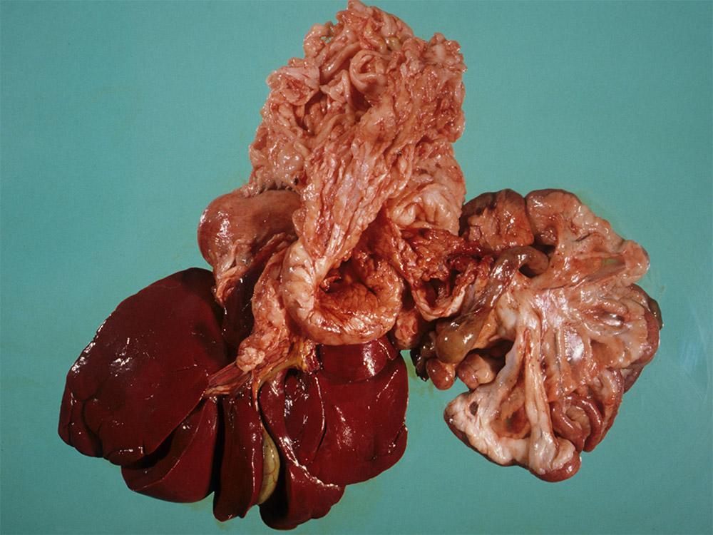
The liver, shown below, is examined initially, before removal of the alimentary tract.
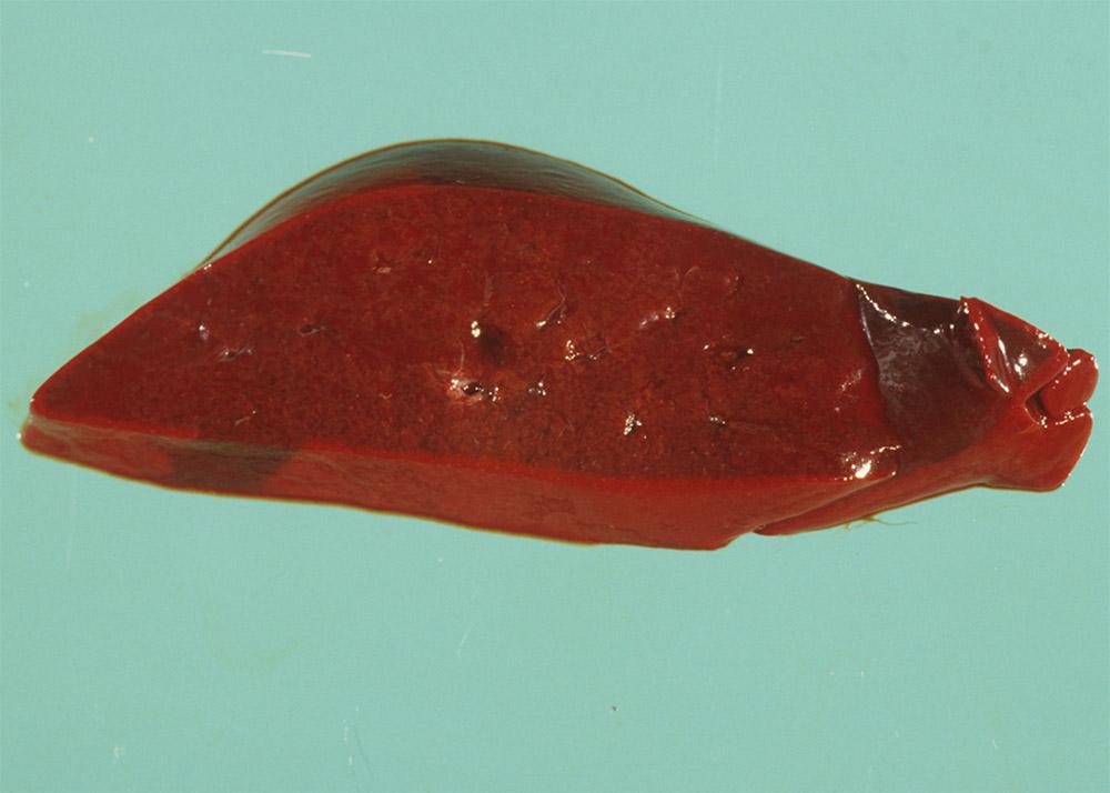
Next, examine the liver, and cut into it to examine the parenchyma.
This cross-section shown below is normal.
Alimentary tract
Stomach
Next, examine the stomach. If the oesophagus is still attached, start by cutting along the oesophagus. If the oesophagus has been removed previously, insert the scissors into the cardia and cut along the lesser curvature (dotted line).
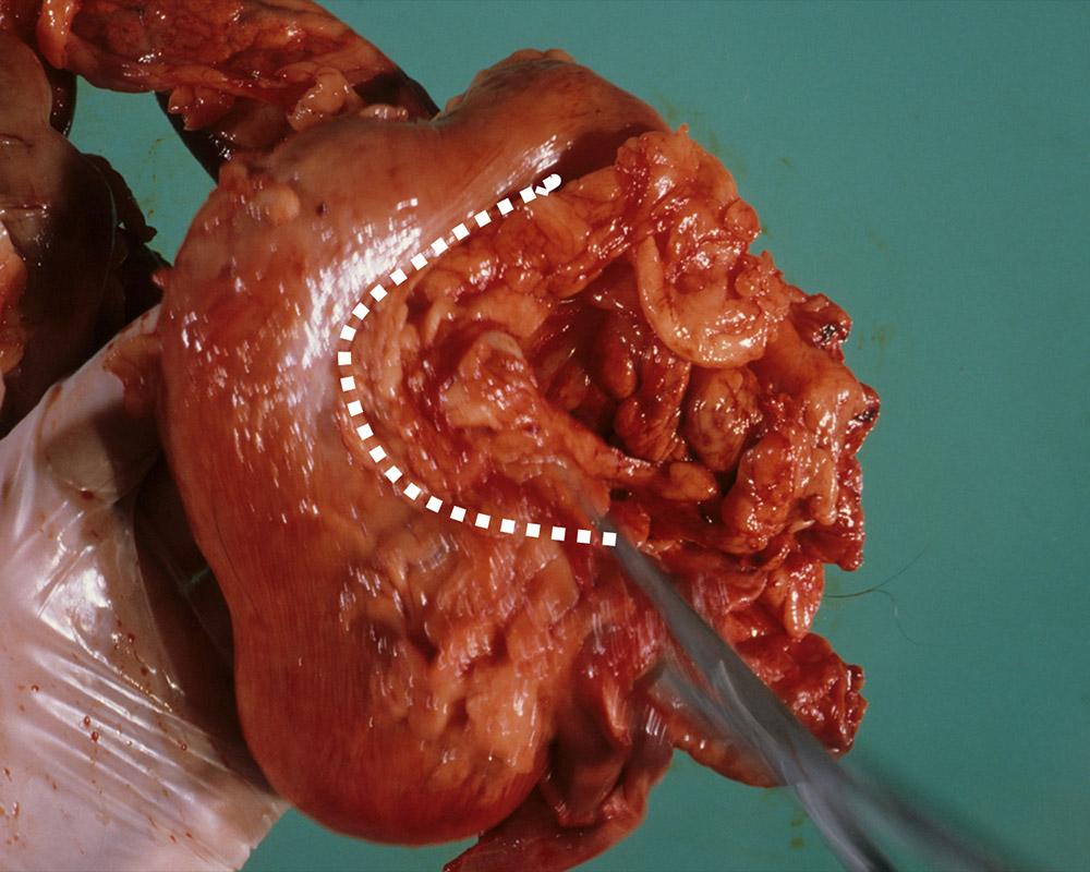
This opens the stomach and allows the mucosa to be examined.
Intestinal tract
Continue by examining the intestinal tract.
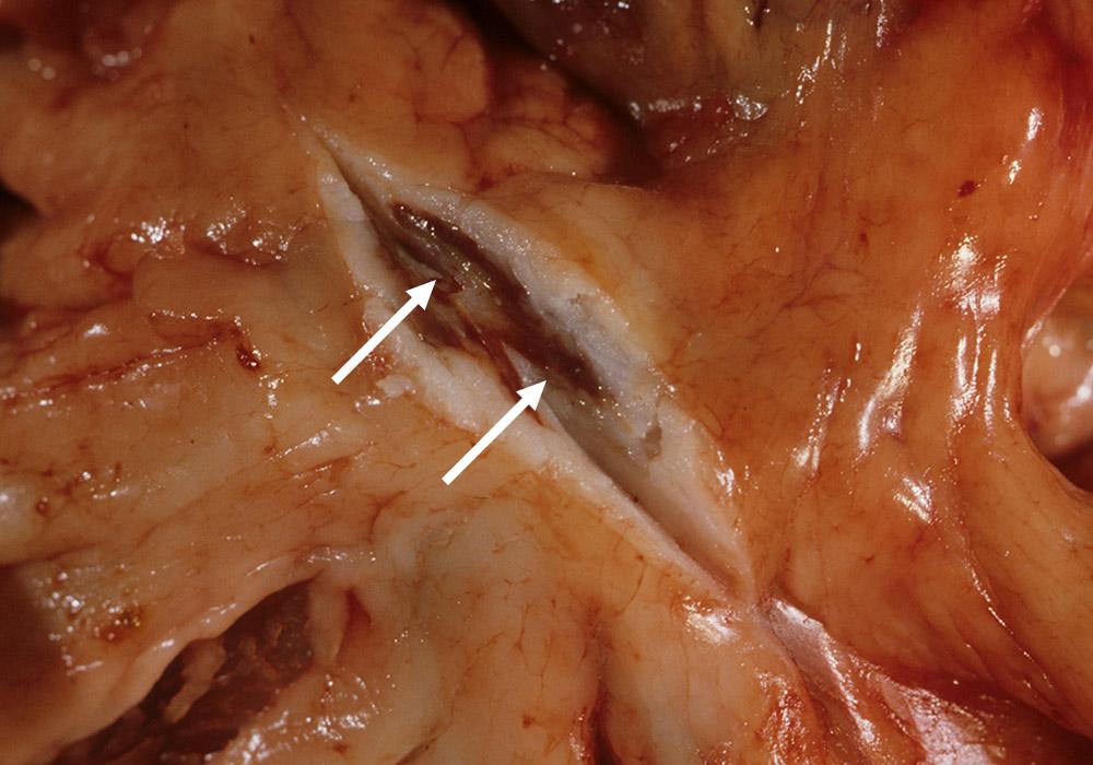
Feel for a small, firm structure in the mesenteric fat, and cut into it with a scalpel to examine its structure. The brown structure in the image below is a normal, mesenteric lymph node (arrows).
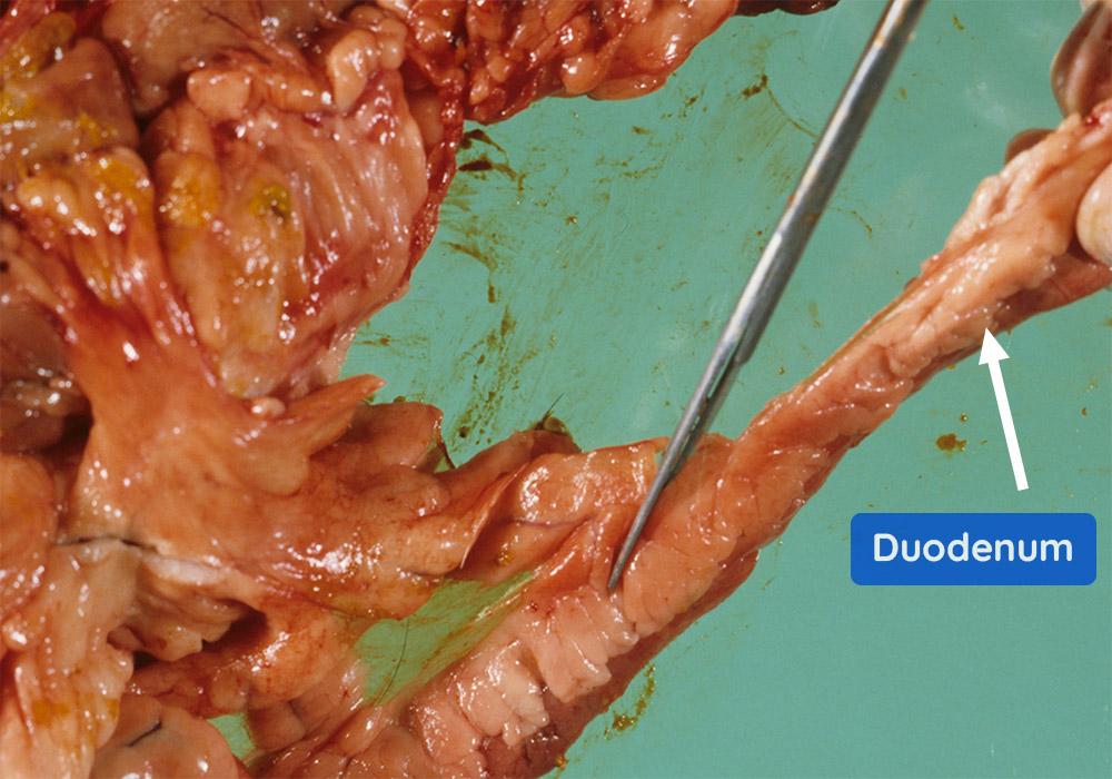
The intestinal tract can be examined ‘in situ’ attached to the mesentery, but is easier if separated first. Hold the intestine at the duodenum and cut along the mesenteric attachment (as shown below) to the ileum.
The alimentary tract can then be laid out as shown below.
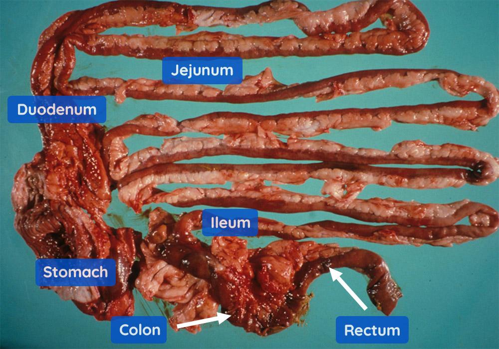
Note: If lesions are suspected in the alimentary tract, samples of stomach, duodenum, jejunum, ileum, colon and rectum should be collected before the alimentary tract is removed. Why? To ensure that they are fixed in 10% neutral-buffered formalin quickly to prevent tissue autolysis (breakdown).
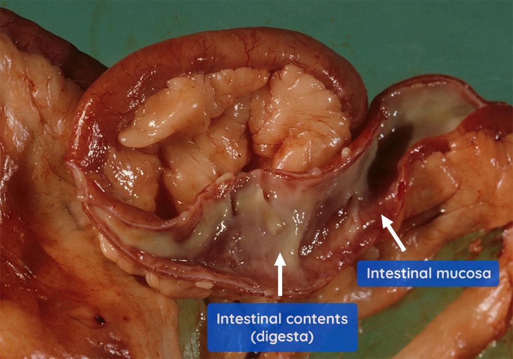
To examine the mucosa and contents, cut the intestine along its antimesenteric edge (see image below). In this case, the contents are creamy grey in colour, which is normal. The mucosa is reddish/brown, which is also normal.
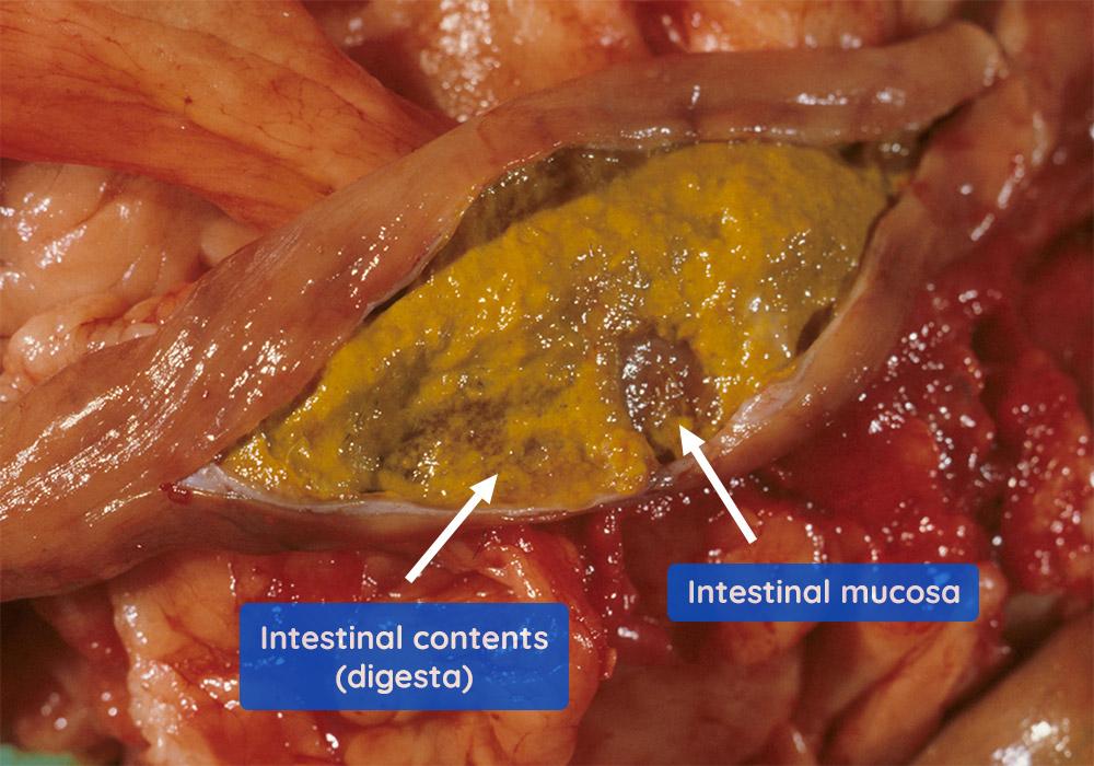
The image below shows the opened colon. The contents are yellow and the mucosa is grey/brown. Both of these are considered normal.
Spleen
Now we will examine the spleen.