

Overview
Epizootic lymphangitis (EZL) is a contagious, chronic disease of horses, mules and donkeys caused by the fungus Histoplasma capsulatum var. farciminosum. Also referred as ‘pseudo-glanders’, this disease is endemic and highly prevalent in North Africa and parts of Asia, namely India, Pakistan and Japan.
Clinical signs
EZL presents in three different forms (cutaneous, ocular or respiratory), likely dependent on the route of entry of the causative agent:
Cutaneous form
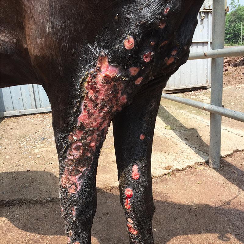
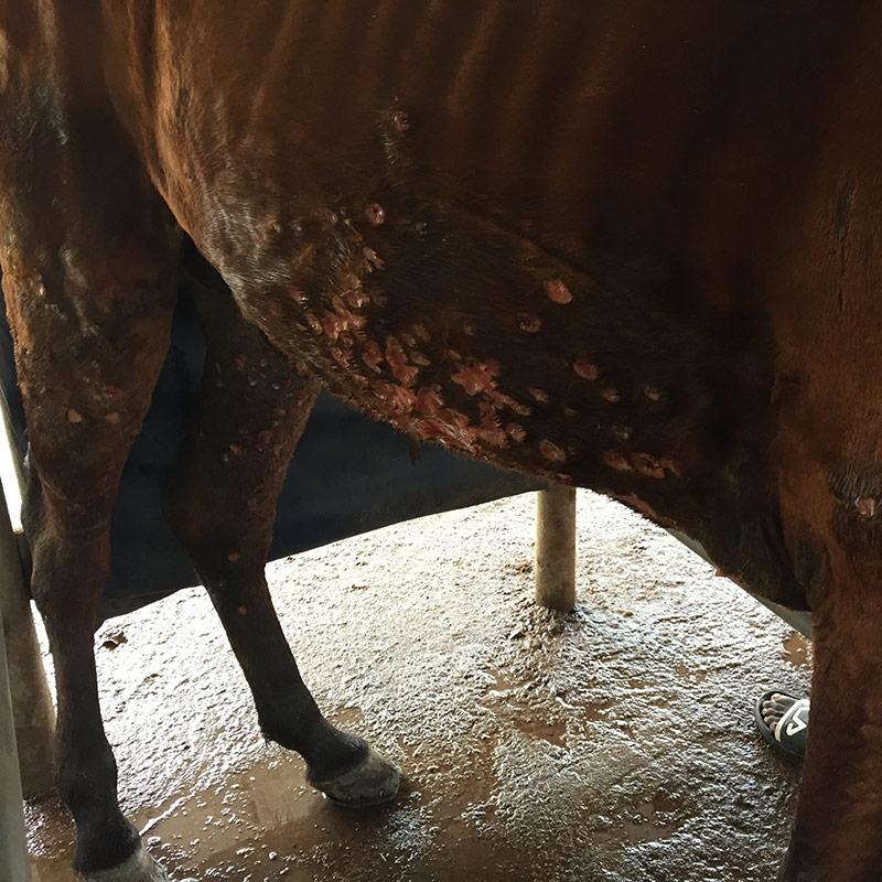
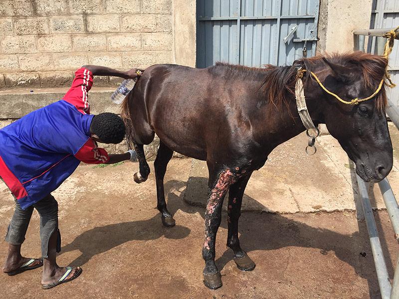
Characterised by suppurative, ulcerating, rapidly spreading pyogranulomatous, multifocal dermatitis and lymphangitis. Commonly affecting the extremities, chest wall and the neck (figure 1).
Ocular form
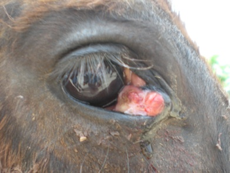
Presenting as a proliferative and exudative conjunctivitis of the palpebral conjunctiva (figure 2).
Respiratory form
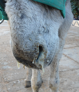
The respiratory form is the most rare and causes respiratory symptoms (figure 3) and multifocal pneumonia.
💡 Whilst Histoplasma spp. are known to cause infection in humans, there are no reports of equine-human transmission of this potentially zoonotic disease
Transmission
Infection occurs directly by contact on open/damaged skin with infected pus, nasal or ocular secretions, or indirectly, by soil or contaminated harnesses, grooming equipment, feeding and watering utensils, wound dressings or flies and ticks.
Flies seem to be the main vector for spreading of the conjunctival form, while the pulmonary form appears to require inhalation of the organism and is therefore less common.
Irrespective of the infection route, the incubation period is from around 3 weeks to 2 months. In all cases, the lesions are nodular and granulomatous in character, and the organism, once established, spreads locally by invasion and then via the lymphatics.
Diagnosis
It is important to detect and diagnose the causative agent as the clinical disease can appear similar to a skin form of glanders known as ‘farcy’, as well as pseudotuberculosis, rhodococcal ulcers, cryptococcosis, strangles, sarcoids and cutaneous lymphosarcomas. While microscopic identification through stained impression smears, culture and hystopathology is possible, as well as a number of serological tests, the organism is often difficult to isolate and identify, and a PCR based test is ultimately preferred.
Treatment
Surgical lancing of skin nodules and/or scab-picking, followed by packing the areas with gauze soaked in 7% tincture of iodine is the main treatment option (figure 4). Intravenous or oral dosing of potassium iodide in a 10% solution given once a week for four weeks is the best option in endemic areas, albeit expensive and difficult to source.
Successful treatment with amphotericin B at a dose of 0.2 mg/kg bodyweight, three times on alternate days, has also been reported, as has griseofulvin combined with iodides and local surgical treatment.
Prevention
Prevention is based on community awareness promoting early detection and intervention. Suspected cases should be isolated and treated, or euthanased if too severe or unresponsive to therapy. In addition, thorough and extensive cleaning and disinfection of the environment and transmission vectors (such as tack, brushes, water/feed throughs/buckets) are required to prevent the disease from spreading.